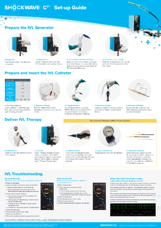
shockwave c2 plus logo

PULSES

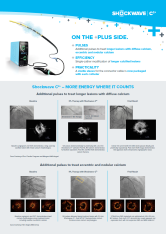
Additional pulses to treat longer lesions with diffuse calcium, eccentric and nodular calcium

EFFICIENCY

Single-catheter modification of diffuse calcium within longer lesions

PRACTICALITY

A sterile sleeve for the connector cable is now packaged with each catheter
Shockwave C2 IVL catheter

The +Plus Side of Coronary IVL

Shockwave C2+: More Energy Where it Counts

Open All Close All

Diffuse Calcium within Longer Lesions

Efficient treatment of diffuse calcium within longer lesions with Shockwave C2+

Case Courtesy of Dr. Nikos Werner Krankenhaus der Barmherzigen Brüder Trier, Germany
Baseline
Guide extension-facilitated delivery of IVL
120 pulses delivered distally to proximally with single 3.0mm Shockwave C2+ catheter treating ~100mm of the lesion
Final result after DES

Nodular Calcium

50% more pulses for challenging calcium morphologies along the treatment lesion with Shockwave C2+

Case Courtesy of Dr. Angela McInerney Cardiovascular Institute, Hospital Clínico San Carlos Madrid, Spain

Baseline

Mixed calcium morphologies along treatment lesion including nodular calcium

IVL Therapy with Shockwave C2+

IVL pulses delivered with 2.5 mm Shockwave C2+. Post IVL OCT demonstrates calcium fracture across calcium morphologies

Final Result

2.75 x 24 mm DES implanted and optimized via 3.0 & 3.5 NC balloon. Post stent OCT demonstrates a well opposed and expanded stent with min expansion 98% and MSA 5.86 mm2

Eccentric Calcium

First-line calcium modification of mixed calcium morphologies with Shockwave C2+

Case Courtesy of Dr. Margaret McEntegart Columbia University Irving Medical Center New York, NY, United States
Case Courtesy of Dr. Claudia Cosgrove St George’s Hospital │ London, UK

Baseline

Baseline angiogram and IVUS demonstrate a long, severely calcified lesion with mixed calcium morphologies.

IVL Therapy with Shockwave C2+

120 pulses delivered distally to proximally with a 3.0 mm Shockwave C2+ catheter. Post IVL NC balloon demonstrates full balloon expansion. Post IVL and NC IVUS demonstrates calcium fracture.

Final Result

3.0 x 32 mm and 3.5 x 38 mm DES stents placed distally and proximally respectively. Post stent IVUS image demonstrates well apposed stent & impressive angiographic result.
Open All Close All

See How Shockwave IVL Performs
Across Calcium Morphologies

360 degrees icon
concentric
180 degrees icon
Eccentric
int coronary nodular
Nodular
Learn More

Shockwave C2+ Specifications

packaging for c2 plus device
DIAMETER (mm) 2.5, 3.0, 3.5, 4.0
LENGTH (mm) 12
GUIDEWIRE COMPATIBILITY (in) 0.014
GUIDE CATHETER COMPATIBILITY 5Fr
WORKING LENGTH (cm) 138
CROSSING PROFILE RANGE* (in) 0.044-0.047
PULSES (max) 120

Working Lengths

Balloon working length of 12mm from inner edge of marker bands

6mm between emitters and 4mm/2mm between emitters and marker bands

Diagram

Energy Profile

Acoustic pressure declines ~3mm away from the emitter

Smooth energy profile over center 12mm of balloon

2
mm
Diagram
Diagram

Optimized Overlap

2mm overlap optimizes energy profile

Shockingly Sound Clinical Evidence

CAD Pooled data demonstrates safety, efficacy and intuitiveness within a global patient population

patients icon
628
Patients
green city icon
72
Sites
pin earth icon
12
Countries
Explore CAD Pooled Data

Shockwave C2+ Resources

Shockwave C2+ Tech Sheet thumbnail
Shockwave C2+ Tech Sheet
Download
ifu portal img
Shockwave IFU Portal
cs plus setup guide
Shockwave C2+
Set Up Poster
Download
Sterile Sleeve Video shot
Covering the Connector Cable with the Sterile Sleeve
WATCH VIDEO

Hear What Users are Saying About Shockwave C2+

Ready to Make Waves with Shockwave Medical?

Looking for careers? Click here.